

Lower bone density in the thoracic spine is associated with faster cognitive decline in adults, according to a study by Johns Hopkins University researchers.
The findings, published in the medical journal Radiology, show that reduced volumetric bone density in the middle back correlates with structural brain changes linked to impaired memory, executive function, and an increased risk of dementia.
An interdisciplinary research team re-analyzed health data from the Multi-Ethnic Study of Atherosclerosis, examining non-contrast chest computed tomography scans from 2,086 participants. The final analysis focused on 715 individuals who had available volumetric bone density measurements as well as brain magnetic resonance imaging scans and cognitive tests.
To determine volumetric bone density in the thoracic vertebrae, the researchers used a deep learning algorithm developed at Johns Hopkins University. The automated tool calculated mineral density directly from routine chest CT scans taken at the start of the study.
Johns Hopkins University, based in Baltimore, Maryland, is a major center for medical research and clinical care. Radiology, published by the Radiological Society of North America, is a leading peer-reviewed journal in diagnostic imaging. Non-contrast computed tomography uses X-rays to capture cross-sectional anatomical images without dye injections, while magnetic resonance imaging uses magnetic fields and radio waves to detail soft brain tissues.
Brain Microstructure and Cognitive Changes
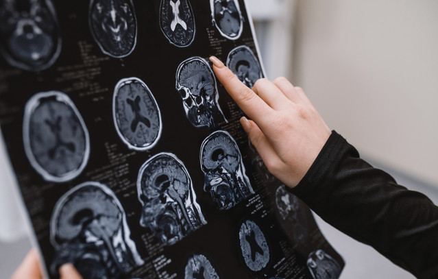
The research team examined brain MRI scans to track specific microstructural alterations, including areas of white matter hyperintensities and reduced fractional anisotropy. Prior medical research links both structural changes to cognitive decline and heightened dementia risk in older adults.
The analysis demonstrated that lower baseline volumetric bone density in the thoracic spine was linked to faster overall cognitive decline. At the structural level, lower bone density correlated with a higher concentration of hyperintense foci in the white matter of the corpus callosum, a region supporting executive functions, working memory, and attention.
Lower spine bone density was also tied to a faster decrease in fractional anisotropy within the anterior limb of the internal capsule, another neural pathway involved in executive functioning. White matter nerve fibers act as communication channels across the central nervous system, with the corpus callosum bridging the left and right brain hemispheres and the internal capsule relaying signals between the cerebral cortex and lower brain centers.
Shared Metabolic Factors in Aging
The study authors emphasized that their findings do not indicate that osteoporosis directly causes dementia. Shadpour Demehri, a professor of radiology at Johns Hopkins University, explained that the study was not designed to show a cause-and-effect relationship, but rather to observe a metabolic syndrome that may simultaneously trigger bone and brain degeneration.
Demehri noted that the co-occurrence of reduced bone density and brain changes likely reflects shared metabolic factors associated with aging. These potential drivers include insulin resistance, dyslipidemia, and hormonal changes during menopause, which can affect both bone mineral maintenance and neural health across time.
Bone mineral density measures the quantity of key minerals, such as calcium, within a given volume of bone tissue. When bone density declines, bones grow weak and fragile, significantly raising the risk of fractures. Osteoporosis and cognitive impairment are two common conditions in aging populations that frequently progress without obvious early symptoms.
Early Detection and Future Prevention
The researchers noted that identifying individuals with low volumetric bone density on routine chest scans could help clinicians spot patients at risk of parallel decline in skeletal and neurological health. Early identification could enable timely screening and targeted management of shared metabolic risk factors before serious degeneration occurs.